X-ray crystallography is a powerful tool in exploring and analyzing biomolecular structures and nanomaterials. But with the rapid development of nanoscience and biology, it has become an urgent need to resolve the internal structure of noncrystalline samples.
Traditional x-ray microscopy methods obtain the image of a noncrystalline sample by recording the intensity of the light beam passing through the sample with a multi-pixel detector. However, it is different in ghost imaging, where the sample’s image is reconstructed via correlations between the intensities of two light beams: one beam hits the sample and is recorded with a single pixel detector, while the other does not hit the sample and is recorded directly. But so far, ghost imaging has only been demonstrated with visible and infrared light.
A research group in the Key Laboratory for Quantum Optics, Shanghai Institute of Optics and Fine Mechanics, Chinese Academy of Sciences, and co-workers from the Shanghai Institute of Applied Physics, Chinese Academy of Sciences, now extend this imaging capability of ghost imaging to the x-ray regime. It is for the first time that the Fourier-transform ghost imaging (FGI) is successfully demonstrated with hard X rays in an experiment.
The experiment was performed on the 13W beamline at the Shanghai Synchrotron Radiation Facility (SSRF). A pseudothermal x-ray source, which can generate controllable chaotic x-ray speckle pattern fluctuations to emulate the behavior of a spatially incoherent x-ray source, was used in the experiment. The x-ray energy was centered at 12.1 keV (0.1nm wavelength). By measuring the second-order intensity correlation function of the light, Fourier-transform diffraction pattern of a complex amplitude sample is achieved at the Fresnel region in the experiment and the amplitude and phase distributions of the sample in the spatial domain are retrieved successfully.
This method extends x-ray crystallography to noncrystalline samples and, as a lensless microscopy scheme, the spatial resolution of x-ray FGI is only limited by the x-ray wavelength in principle. An important feature of the x-ray FGI method is that it does not rely on a highly coherent source. Therefore, it provides a feasible way to achieve high resolution images of noncrystalline samples with widely accessible laboratory x-ray sources.
In addition, eliminating the need for an intensive coherent source, it also provides a glimpse of the possibility of revolutionizing the current neutron and electron scattering methods widely used in research of materials science as well as biomedicine.
Their research was published in journal of Physical Review Letters with title Fourier-Transform Ghost Imaging with Hard X Rays.
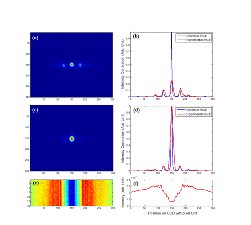
FIG. 1. Diffraction patterns of the sample obtained with X-rays of 0.1nm wavelength. (a) is the Fourier-transform diffraction pattern of the sample’s transmittance obtained by X-ray FGI, (c) is the Fourier-transform diffraction pattern of the squared modulus of the sample’s transmittance obtained in X-ray FGI. The red lines in (b) and (d) are the cross-section curves of (a) and (c), respectively. The blue lines in (b) and (d) are the corresponding theoretical results. Image by YU Hong
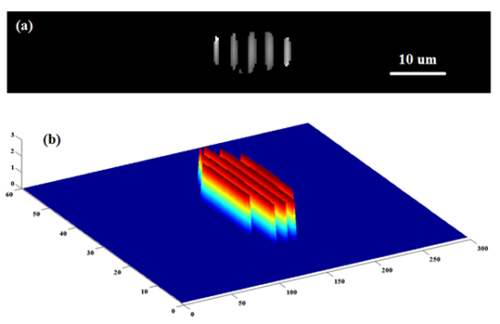
FIG. 2. The sample’s distributions in spatial domain retrieved from the Fourier-transform diffraction patterns obtained in X-ray FGI. (a) and (b) are the amplitude and phase distributions of the sample’s transmittance. The pixel size in the retrieved image is 0.297um. Image by YU Hong
