Chinese research team has developed an advanced imaging technique to achieve super-resolution microscopy at unprecedented speeds and with many fewer images. The new method should make it possible to capture processes in living cells at speeds not previously possible.
Super-resolution techniques, often called nanoscopy, achieve nanoscale resolution by overcoming the diffraction limit of light. Although nanoscopy can capture images of individual molecules inside cells, it is difficult to use with living cells because hundreds or thousands of images are needed to reconstruct an image -- a process too slow to capture quickly changing dynamics.
In Optica, a journal for high impact research published by The Optical Society (OSA),investigators from Shanghai Advanced Research Institute of the Chinese Academy of Sciences and the Shanghai Institute of Optics and Fine Mechanics of the Chinese Academy of Sciences described how they used an unconventional imaging approach known as ghost imaging to enhance the imaging speed of nanoscopy. Their new technique produces nanometer resolution using orders of magnitude fewer images than traditional nanoscopy techniques.
"Our imaging method can potentially probe dynamics occurring on millisecond time-scales in subcellular structures with spatial resolution of tens of nanometers - the spatial and temporal resolution at which biological processes take place," said WANG Zhongyang, co-leader of the research team.
The new approach is based on stochastic optical reconstruction microscopy (STORM), one of three researchers sharing the Nobel Prize in Chemistry in 2014. STORM, which is also sometimes called photoactivated localization microscopy (PALM), is a wide-field technique that uses fluorescent labels that switch between light-emitting (on) and dark (off) states.
Acquiring hundreds or thousands of images, each capturing the subset of fluorescent labels that are on at a given time, allows the location of each molecule to be determined and used to reconstruct a fluorescence image.
The researchers turned to ghost imaging to speed up the STORM imaging process. Ghost imaging forms a picture by correlating a light pattern that interacts with the object with a reference pattern that does not.
Individually, the light patterns don't carry any meaningful information about the object. The researchers also used compressive imaging, a computational approach that enables image reconstruction with fewer exposures because it uses an algorithm to fill in the missing information.
"While STORM requires a low density of fluorescent labels and many image frames, our approach can create a high-resolution image using very few frames and a high density of fluorophores," said one of the research team's other co-leaders, HAN Shensheng. "It also doesn't need any complex illumination, which helps reduce photobleaching and phototoxicity that could harm dynamic biological processes and living cells."
To implement the new technique, the researchers used an optical component known as a random phase modulator to turn fluorescence from the sample into a random speckle pattern. Coding the fluorescence in this way allowed each pixel of a very fast CMOS camera to collect light intensity from the whole object in a single frame.
To form the image via ghost imaging and compressive imaging, the light intensity was correlated with a reference light pattern in a single step. The result was more efficient image acquisition and a reduction in the number of frames required to form a high-resolution image.
The researchers tested the technique by using it to image a 60-nanometer ring. The new nanoscopy approach resolved the ring using just 10 images while traditional STORM approaches would have needed up to 4000 frames to achieve the same result. The new approach also resolved a 40-nanometer ruler with 100 images.
"We hope this method can be applied to a variety of fluorescent samples, including those that exhibit weaker fluorescence than those used in this research," said WANG.
The researchers also want to make the technique faster to achieve video-rate imaging with a large field of view in order to acquire 3D and color images.
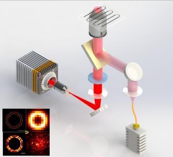
Nanoscopy Setup(Image by SIOM)
Article website:
https://doi.org/10.1364/OPTICA.6.001515
Contact:
Mr. CAO Yong
General Administrative Office
Shanghai Institute of Optics and Fine Mechanics, CAS
Email: caoyong@siom.ac.cn
Source: EurekAlert 12-Dec-2019
Home | About SIOM | Newsroom | International | Admissions | Publications | Contact
@ Shanghai Institute of Optics and Fine Mechanics Tel:02169918000 沪ICP备05015387号-1 Technical support:Qingyun Software