As a newly developed coherent diffraction-imaging (CDI) imaging method, the ptychographical iterative engine (PIE) not only can bypass the difficulty of having high-quality optics in x-ray microscopy by a numerical reconstruction algorithm, but also has obvious advantages on traditional CDI methods in both converging speeds and view fields. However, like in the other CDI methods, the reconstruction of the image from the intensity data of a weakly diffracting specimen is still difficult because of the low signal to noise ratio. To improve this situation,researchers from Prof. Zhu Jianqiang’s group at Shanghai Institute of Optics and Fine Mechanics, Chinese Academy of Sciences(SIOM, CAS) reported an optical setup for the data recording and a corresponding algorithm for the image reconstruction. The result of their work explains a phenomenon that has confused the researchers for many years: even though the CDI imaging theory requires using the square root of the recorded data, in practical experiments of x-ray imaging, a reconstruction with 12(I(k))n' type="#_x0000_t75"> can have better contrast for some cases. [Optics Letters, 37(16), 3348-3350 (2012).].
They suggest the use of divergent light beams for the illumination in the data recording process and a modified ptychographical iterative engine (PIE) algorithm for the image reconstruction to get a remarkable contrast enhancement. The results obtained can also be extended to other CDI techniques. The optical setup for the PIE technique is schematically shown in Fig. 1.
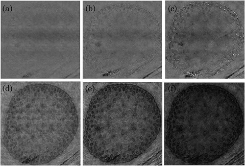
The validity of the above analysis is verified by a numerical simulation and experiment. Fig. 2 is the numerical simulation result. The experimental results are shown in Fig. 3.

Fig. 1. Schematic diagram of the optical setup.

Fig. 2. Numerical simulation results. (a) The phase transmission of the object; (b) the diffraction pattern; (c) the reconstructed image with common PIE method; (d) the reconstructed image with they suggested method.

Fig. 3. Experimental results. (a)–(c) Phase and (d)–(f) corresponding intensity imagereconstructions with,![]() , respectively
, respectively